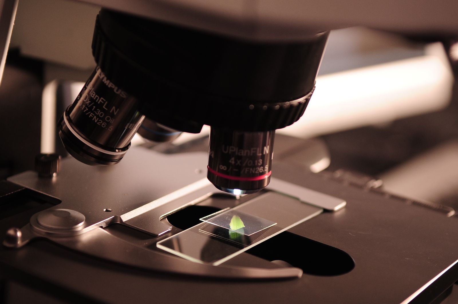
Under a compound microscope in London in 1665, Robert Hooke pressed a thin shaving of cork against the stage, angled a candle through a water-filled glass sphere to concentrate the light, and looked down at what he later described as a honeycomb of tiny empty rooms. He counted them in great numbers throughout the cork. He needed a word for the little chambers. He called them cells, after the bare rooms monks slept in inside a monastery. The name stuck. Every heart, every leaf, every bacterium, every embryo on Earth is now measured in the unit he named that afternoon.
The observation appeared in Micrographia, the illustrated folio Hooke published later that same year, which became a widely celebrated scientific publication. Samuel Pepys read it enthusiastically and wrote in his diary that it was the most ingenious book he had ever seen.
A sliver of bark, sliced with a penknife
The specimen was ordinary. Cork is the outer bark of the cork oak, Quercus suber, and by the 1660s it was already a common stopper for wine bottles and apothecary jars across Europe. Hooke, working at the Royal Society, wanted to know why it was so light and springy. He took a clean piece, sharpened a penknife “as keen as a razor,” and shaved off a slice thin enough for light to pass through.
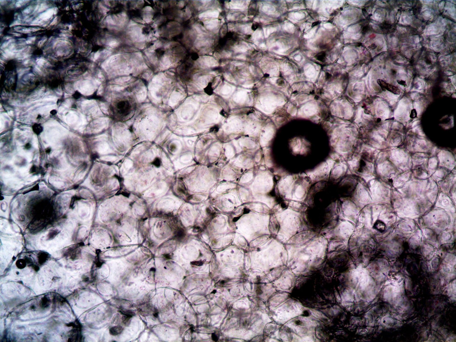
What he saw through his microscope was structure. The cork was not solid at all. It was a lattice of hollow polygonal boxes, walled off from each other, arranged in tidy rows. The walls were extremely thin. The emptiness explained the buoyancy. It also gave him the metaphor.
“These pores, or cells,” Hooke wrote, “were not very deep, but consisted of a great many little Boxes.” Science Friday traces the naming directly to the Latin cella, the word for a small chamber, the same root that gives us the cells in a prison and the cells a monk retreated to for prayer. Hooke chose the word because the compartments were empty and identical, like the rooms in a religious dormitory. He was describing dead plant tissue. The cell walls were the ghosts of once-living structures. He never saw a living cell in cork, because cork is what remains after the cells inside have died and their contents drained away.
The instrument on the table
The microscope Hooke used was a compound instrument, meaning it had two lenses stacked in a tube — an objective at the specimen end and an eyepiece at the viewer’s end — which multiplied their magnifying power. The instrument was carefully constructed, mounted on a pillar so Hooke could tilt it. Beside it sat an oil lamp, its flame focused through a water-filled globe that acted as a condenser lens to throw a bright, even light onto the specimen.
Magnification was modest by modern standards. The image was blurred by chromatic aberration — the coloured fringes that plague simple glass lenses — and Hooke often had to compare what he saw through the compound scope with what he saw through a single, higher-power lens to check that he was not being tricked by an optical artifact. The evolution of glass lenses over three centuries shows how much of early microscopy was really a story about glassmakers. Better lenses meant better biology.
Why cork, and why then
Cork was not a random pick. Hooke was working through a menagerie of everyday objects — the point of a needle, the edge of a razor, the sting of a bee, a flea, a louse, the eye of a fly. He wanted to know what familiar things looked like when magnified past the threshold of unaided vision. Cork happened to be the specimen where the visual grammar of biology first snapped into focus.
The timing mattered too. Compound microscopes had existed for several decades by 1665, but nobody had systematically turned one on living matter and published what they saw. Hooke’s Micrographia was the first sustained tour of that hidden world. A Nature collection on four hundred years of cork imaging traces how every subsequent characterisation of cork’s cellular structure builds on the observation Hooke made that year.
From empty boxes to a theory of life
Hooke’s cells were, in a strict sense, misleading. They were empty. He had found the walls, not the living contents. It would take time for scientists to grasp that every plant and animal is built from these units and that the units are alive — sacs of watery chemistry, not hollow rooms.
That leap came in the 1830s, when botanist Matthias Schleiden and physiologist Theodor Schwann proposed what became known as cell theory: that all living tissue, plant or animal, is composed of cells as its fundamental units. Rudolf Virchow added the third pillar in the mid-19th century — omnis cellula e cellula, every cell comes from another cell. The empty chambers Hooke had counted in cork bark turned out to be the same architectural unit that made up muscle, nerve, blood, bone, leaf, root, and every microbe on the planet.
By the 21st century, the cells in a single human body are estimated at roughly 37 trillion. MIT Technology Review profiled Aviv Regev, the computational biologist co-chairing the Human Cell Atlas, an international project cataloguing every cell type in the human body. Regev has described the finished atlas using the metaphor of a periodic table for cells. Lander has compared the expected impact of the cell atlas to that of the Human Genome Project, suggesting it will be similarly transformative.
The vocabulary is Hooke’s. The unit is Hooke’s. The instrument has been replaced many times over, but the word has not.
What Hooke could not see
The cell walls of cork are made of a waxy polymer called suberin, deposited by the tree to waterproof its outer bark. Hooke could not have known this. He had no chemistry to speak of, no stains, no fluorescent dyes. He was working with white light, glass, and geometry.
Modern micrographs of the same tissue reveal a scaffolding of chambers packed with structure — membranes, protein machines, coiled DNA. The trippy pinks and neon greens of contemporary cell imagery are, as Jack Challoner explains in a book Hyperallergic describes as a 21st-century update of Micrographia, added by scientists using contrast, fluorescence, and stain. Challoner explained to the magazine that cellular structures lack natural color, requiring scientists to add contrast and fluorescence. Challoner noted that electron microscopy cannot capture color. The dazzling images are the modern equivalent of Hooke’s engravings: technique-heavy renderings that translate the invisible into something a human eye can read.
What survives from 1665 is the concept. A cell is a bounded space that does something. Everything else — the membranes, the organelles, the ribosomes, the mitochondria, the double helix at the centre — is upholstery inside Hooke’s monk’s room.
The cell as unit of everything else
Once biology accepted that all life is cellular, every downstream question inherited the framing. When Alfred Hershey and Martha Chase conducted their famous blender experiment in 1952 to work out whether genes were made of DNA or protein, the whole design assumed they were tracking molecules moving in and out of bacterial cells. The Cold Spring Harbor experiment settled that DNA carries genetic information — a discovery only intelligible because Hooke’s unit was already the accepted vocabulary of life.
The same is true for the strangest edge cases of biology. A tiny hydrozoan called Turritopsis dohrnii can, when injured, reverse the differentiation of its adult cells and revert to a juvenile polyp, effectively restarting its life cycle. The trick is a cell-level trick. Nothing about that story is describable without Hooke’s word.
Cancer is a cell-level failure. Antibiotics are cell-level warfare. Vaccines are cell-level training. mRNA therapies deliver instructions to individual cells. CRISPR edits the genome inside single cells. The entire toolkit of 21st-century medicine operates at the scale Hooke first named.
The book that made the microscope famous
Micrographia ran to more than sixty engravings, several of them fold-outs a metre wide. The flea plate was the most famous — a monstrous armoured insect rendered in exquisite detail across two pages, its bristled legs and segmented body magnified to the size of a small dog. Readers had never seen anything like it. Hooke’s 1665 flea illustration remains one of the defining images of what a microscope revealed to a startled public.
Pepys called it “the most ingenious book that I ever read in my life.” Isaac Newton, then a student at Cambridge, bought a copy and made notes in the margins. It sold out. Hooke’s engravings did for microscopy what the Hubble Space Telescope’s deep-field images would do for astronomy three centuries later: they turned an obscure instrument into a public event.
Hooke himself was a man of near-manic range. In addition to running the microscopy programme at the Royal Society, he surveyed London after the Great Fire of 1666, designed buildings, argued with Newton about gravity, formulated the law of elasticity that still bears his name, and coined a technical vocabulary that biology still uses. His portrait, if one ever existed, has been lost. What remains is the word.
The monks’ rooms, four centuries on
A cork oak in Portugal today, its bark harvested every nine years for the wine industry, is producing the same waxy chambers Hooke drew in 1665. The trees can live for two hundred years. Some cork oaks currently being stripped for stoppers were seedlings when Micrographia was still in print.
Look at a wine bottle. The stopper in the neck is Hooke’s specimen — dead plant tissue, walled off in polygonal compartments, still holding the pattern he described by candlelight. The word for those compartments has travelled from a London workshop through cell theory, germ theory, genetics, molecular biology, and the Human Cell Atlas. It has been applied to bacteria that live in boiling vents, to neurons in the cortex, to the fertilised egg that becomes a human, to the T-cell hunting a virus in the bloodstream.
Hooke needed a word for empty rooms. He borrowed one from the monasteries. Every biology textbook on Earth still uses it.